
Videos
Problem Set
QUANTITATIVE Limits of Resolution Then and Now. Based on what you learned in this chapter about the limit of resolution of a light microscope, answer each of the following questions. Assume that the unaided human eye has a limit of resolution of about 0.25 mm and that a modern light microscope has a useful magnification of about 1000×.
(a) Define limit of resolution in your own words. What was the limit of resolution of Hooke’s microscope? What about van Leeuwenhoek’s microscope?
(b) What are the approximate dimensions of the smallest structure that Hooke would have been able to observe with his microscope? Would he have been able to see any of the structures shown in Figure 1-3a? If so, which ones? And if not, why not?
(c) What are the approximate dimensions of the smallest structure that van Leeuwenhoek would have been able to observe with his microscope? Would he have been able to see any of the structures shown in Figure 1-3a? If so, which ones? And if not, why not?
(d) What are the approximate dimensions of the smallest structure that a contemporary cell biologist should be able to observe with a modern light microscope?
(e) Consider the eight structures shown in Figure 1-3a and 1-3b. Which of these structures would both Hooke and van Leeuwenhoek have been able to see with their respective microscopes? Which, if any, would van Leeuwenhoek have been able to see that Hooke could not? Explain your reasoning. Which, if any, that neither Hooke nor van Leeuwenhoek could see would a contemporary cell biologist be able to see using a modern light microscope?
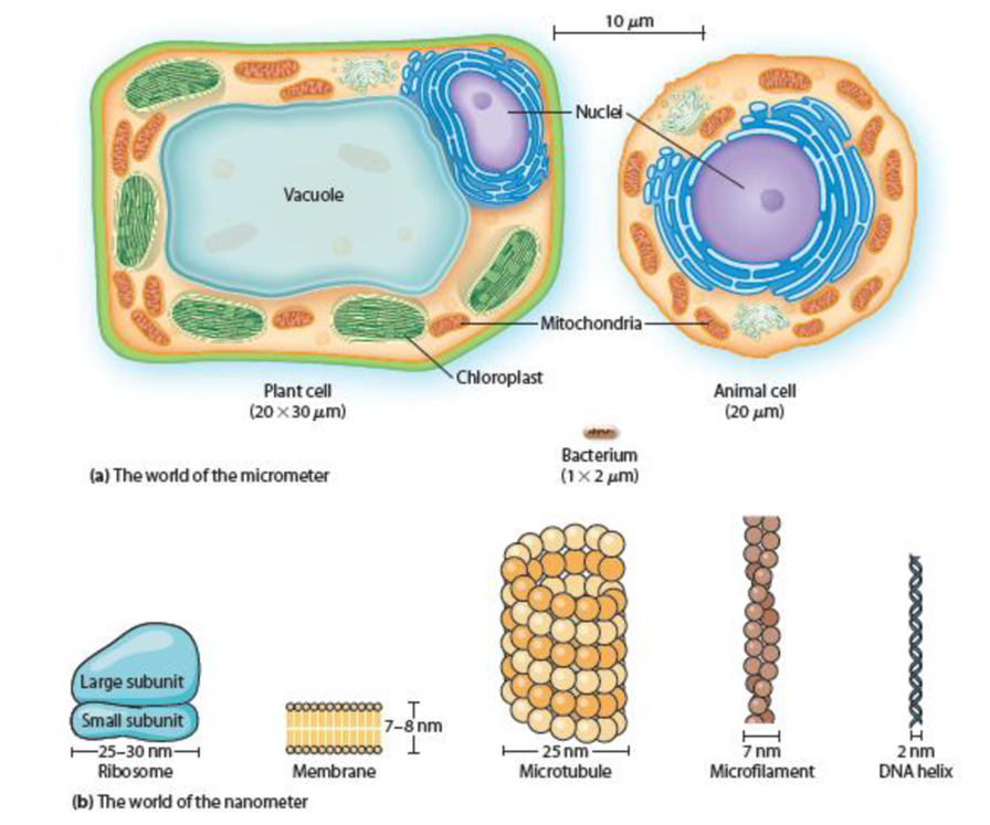
Figure 1-3 The Worlds of the Micrometer and Nanometer. Illustrations show (a) typical cells and (b) common cellular structures.
Want to see the full answer?
Check out a sample textbook solution
Chapter 1 Solutions
Becker's World of the Cell (9th Edition)


- Simulated data: Imagine you collect the following images at the above magnifications. Figure 3-2: Images of cheek cells at the above magnifications. Labelling Drawing: Label only the microscope image taken at 1000x magnification. Add lines and labels (as per guidelines for scientific drawings). You should include: A scale bar showing a unit length of 20 µm. (Hint: The length of the human cheek cell shown is 60 um). Labels for the cell membrane, nucleus, and cytoplasm.arrow_forwardQuestion: The students have some wool samples that they have dyed. They want to find out how much of the dye washes out of the wool at different temperatures. They decide to use colorimetry. Describe a suitable method for this experiment. Include the control variables in your answer. Im stuck answering this question, pls help.arrow_forwardPrevious question: The field of view is the maximum area visible through the lenses of a microscope, and it is represented by a diameter. To determine the diameter of your field of view, you can place a transparent metric ruler under the low power (10X) objective of a microscope. You would focus the microscope on the scale of the ruler and measure the diameter of the field of vision in millimeters. Refer to the image taken of this preparation below. What is the approximate field of view as seen by this 10X objective. Note: This microscope has a 10X eyepiece, giving a total magnification of 100X. The image shows the 1mm is for this question. - Answer for this question was 3mm.arrow_forward
- True or false? each objective of a phase contrast microscope contains a phase plate. most cultured mammalian cells grow best at 37C. the oculars on the compound light microscope are typically 10X.arrow_forward3D dimensionality is a limitation of the compound microscope. Depth of field, DOF, describes dimensionality form top to bottom and can be observed with colored cross threads. Observe the crossed thread slide on low power (4x), then on medium power (10x), then on high power (40x objective magnification). Which crossed fiber is on top? How do you know?arrow_forwardquestion: Can you summarize and explain for me what you want to tell in the article below? Can you explain the figure? When I read it myself, I do not understand exactly what is meant by the article. It would be nice if you could highlight the important points. You can use them in a figure or diagram to explain. thank you and hava a nice day :) Article: Nanotechnology Tools to Inactivate SARS-CoV-2 in Different Environments Outside the Patient SARS-CoV is highly stable at room temperature and at 4 °C, but it is inactivated by ultraviolet light at 254 nm, highly alkaline or acidic conditions of pH >12 or pH <3, respectively, or by brief (e.g., 5 min) heat treatment at 65 °C. SARS-CoV-2 is expected to be similarly sensitive. Several human coronaviruses can be inactivated by classical disinfectants, including bleach, ethanol, povidone-iodine, chloroxylenol, chlorheximide, and benzalkonium chloride, so we expect similar inactivation with SARS-CoV-2. The virus stability on surfaces…arrow_forward
- Question:- calculate the distance between two ocular micrometer division markers in micrometers (μm) when viewed at 100x total magnification, then at 400x.arrow_forwardYou may want to use this resource for this problem. If you do, submit the output along with your solution.You have been given a confocal microscope equipped with the following lasers, excitation filters, andemission filters:Laser Emission filter355 nm 410-470 nm405 nm 470-500 nm488 nm 500-550 nm532 nm 570-610 nm561 nm 610-650 nm640 nm 660-700 nm808 nm 720-780 nmYour task is to design an experiment to visualize the following:1. Nuclei2. A fluorescent protein in the cytosol3. A cell membrane marker antibody conjugated with a fluorophore4. Actin filaments5. LysosomesYou may choose from the following fluorophores for each of the five channels:Nuclei Fluorescent protein Membrane marker Actin marker Lysosome trackerDAPI GFP FITC AF488 Phalloidin LysoTracker RedHoechst 33342 YFP WGA-TRITC AF568 Phalloidin LysoTracker DeepRedSYTO Deep Red RFP Cy7 AF594 Phalloidin LysoTracker Blue Part 3.1Choose appropriate fluorophores for each of the subcellular structures to be imaged, taking into…arrow_forwardPART C: CALCULATING THE DIAMETER OF THE FIELD OF VIEW (FOV)_ The field of view (FOV) is the circular area you can see when you look through the microscope. The diameter of the field of view is different depending on which objective lens you are using. For example, you are using the medium-power objective lens, then the area you can see is actually smaller than if you were using the low-power objective lens. Knowing The diameter of the field of view can help you estimate actual size of objects / cells seen through the microscope. When the revolving nose piece is turned to the low power objective lens, a dear plastic ruler can be placed on the microscope stage (see figure 1). Then, the coarse adjustment knob can be used to focus on the millimeter marks of the ruler making sure that one of the milimeter marks is at the left edge of the field of view (see figure 2). NOTE: Slage cip I-1000 Objects in the FOV are usually measured in micrometers (um). To convert, a FOV in mm, times it by 1000…arrow_forward
- Exercise 2B (optional) - Field of View and Estimation of Size Calculate the diameter of the field of view for each total magnification on your microscope in millimeters (mm) and then convert this value to micrometers (um): 4.5 4,500 um Scanning (40X): 1.8 mm x 100X/40X = mm = Low power (100X): FOV diameter = 1.8 mm = 1800 um High power (400X): 1.8 mm x 100X/400X = 0.45 mm = 450 um =Oal8 mm = 180 Oil immersion (1000X): 1.8 mm x 100x/1000X = um Draw and estimate the length of a single Euglena (high power) and Paramecium (low power): Paramecium Euglena total magnification total magnification FOV diam. um FOV diam. um length um length umarrow_forwardMATERIAL METHOD RESULT SUMMARY Lecia DCM3D confocal Microscope 1) Straumann (Straumann AG, Waldenburg, Switzer- land) 2.2mm in diameter (for short, A) (2) Nobel Biocare (Nobel Biocare AB, Goteborg, Sweden) 2.0mm in diameter (for short, B) Xive Implant System (Friadent GmbH, Mannheim, Germany) 2.0mm in diameter (for short, C) (4) Global D (French) 2.5mm in diameter (for short, D) (5) Sweden & Martina (Padova, Italy) 2.5mm in diameter (for short, E) The microstructural parameters used by the discriminatory analysis revealed many differences between the five manufactures in terms of roughness of the drilling surface. Marenzi et al (2018) have considered that Surface micromorphology, which affects the contact between the drill and bone, has been shown to contribute to heat, wear, and corrosion phenomena due to material abrasion and corrosion resistance through several surface texture indicators Can be seen as a factor. Not only the design, but also the…arrow_forwardDiscussion Give the main differences between the types of microscopes used to diagnose surfaces?) What is the appropriate device to diagnose each of the following and why? O A- The aluminum plate is coated with a nano-solution. b- A colloidal sample containing nano-solutions. C- Study of the microstructure of the surface of a sample of iron.' Is it possible to use an optical microscope to study painted metal) surfaces? And why?arrow_forward
 Human Anatomy & Physiology (11th Edition)BiologyISBN:9780134580999Author:Elaine N. Marieb, Katja N. HoehnPublisher:PEARSON
 Biology 2eBiologyISBN:9781947172517Author:Matthew Douglas, Jung Choi, Mary Ann ClarkPublisher:OpenStax
 Anatomy & PhysiologyBiologyISBN:9781259398629Author:McKinley, Michael P., O'loughlin, Valerie Dean, Bidle, Theresa StouterPublisher:Mcgraw Hill Education,
 Molecular Biology of the Cell (Sixth Edition)BiologyISBN:9780815344322Author:Bruce Alberts, Alexander D. Johnson, Julian Lewis, David Morgan, Martin Raff, Keith Roberts, Peter WalterPublisher:W. W. Norton & Company
 Laboratory Manual For Human Anatomy & PhysiologyBiologyISBN:9781260159363Author:Martin, Terry R., Prentice-craver, CynthiaPublisher:McGraw-Hill Publishing Co.
 Inquiry Into Life (16th Edition)BiologyISBN:9781260231700Author:Sylvia S. Mader, Michael WindelspechtPublisher:McGraw Hill Education