
Concept explainers
Videos
Core Skill: Connections In what other organ is a brush border found (refer back to Figure 46.6)? What general conclusions can you draw about the function of such specialized epithelia?
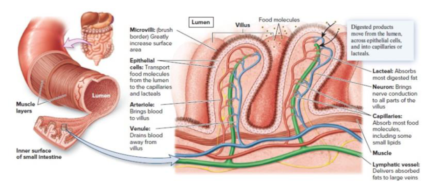
Figure 46.6 The specialized arrangement of tissues in the small intestine. The inner surface of the small intestine is folded into numerous villi, which increase the surface area for digestion and absorption. Within each villus are capillaries and a lymphatic vessel called a lacteal, into which absorbed nutrients are transported. The epithelial cells of the villi have extensions from their surface called microvilli. The microvilli constitute the brush border of the intestine and greatly add to the total surface area.
Want to see the full answer?
Check out a sample textbook solution
Chapter 49 Solutions
Biology
Additional Science Textbook Solutions
Anatomy & Physiology
HUMAN ANATOMY
Human Anatomy & Physiology
Campbell Essential Biology with Physiology (6th Edition)
Evolutionary Analysis (5th Edition)


- Biology / Human Histology *Refer to the attached photo and answer this question: What is the function of the tissue pointed by the arrow?arrow_forwardACTIVITY 1.2: Four Pics One Word Directions: Identify the words that best describe the following pictures. Make a short discussion about the uncovered words.arrow_forwardMultiple Tissues Make Organs You are a part of a research group who wants to build and organ in the lab for organ transplant patients. If this worked the patients don't have to wait for donors. The organ you are building requires the following functional abilities: 1. It has to be able to resist mechanical stresses such as friction and abrasion. This tissue will get sloughed off. 2. The ability to contract involuntarily (you don't have to think about it for it to contract) when stimulated by the nervous system. 3. There has to be a tissue that can stimulate the contraction of the other tissue involved. 4. The ability to resist stress, tension and force from many different directions. 5. There has to be a tissue that acts like glue holding all of the other tissues together to make the organ. Part 1 - Questions you will answer. 1. What are the five tissues that will make up this organ? Justify your choices by describing the characteristics of each known tissue. 2. What organ in the human…arrow_forward
- The structure of a tissue usually is optimized for its function. Describe how the structure of individual cells and tissue arrangement of the intestine lining matches its main function, to absorb nutrientsarrow_forwardIdentifying Main Idea and details. Did you know your second largest organ in your body?arrow_forwardCompare and contrast the characteristics of the ideal tissue forming the outer surface of the body with that forming the lining of the small intestine. How are they similar and different? What tissues fulfill these needs?arrow_forward
- A 10-year-old boy undergoes an appendectomy. Granulation tissue develops normally at the incision site. Tissue remodeling begins at this site with degradation of collagen in the extracellular matrix by which of the following proteins? (A) Cytokines (B) Lipoxygenases C Metalloproteinase (D) Nitric oxide (E) Plasminogen activatorarrow_forwardK MICROSCOPY • EPITHELIAL TISSUE HISTOLOGY INTRODUCTION LABORATORY SIMULATION Labels PHASE 2: Identify str Apical surface types of sir tissue Basal surface Complete the Cell membranes Identify stru simple epith 1 Cell nuclei Cuboidal epithelial cells Lumenarrow_forwardFUNCTION Move fluid that contains foreign over free surface LOCATION Pseudostratified columnar Nasal cavity, lungs, trachea SPECIFIC TYPE MAJOR TISSUE Lines uterus, digestive tract organs Secretion and Kidney tubules, glands and their ducts absorption in the kidney Diffusion, filtration Simple cuboidal Simple squamous epithelium Lining of blood and lymphatic vessels and small ducts Stratified squamous Mouth, throat, and vagina Protection against abrasion Mammary gland ducts, sweat glands, pancreas Male gentilia, pharynx trans Urinary tract Specialized to become distended Loose (areolar) tissue (adipose) skin, hypodermis Loose packing material in most organs Tendons, ligaments words Engish (United States) Skin KFocus G Warrow_forward
- Tissues in a pork chop, make a simple sketch and label the tissues. The bone is a connective layer and supportive tissue the, the lean meat is mostly made up of muscular tissue, the fat is another type of tissue, the white translucent plastic-like material that connects the bone to the lean meat, the lean meatto the fat and the fat to the skin is another type of connective and supportive tissue called the fibrous tissue. plssss help me sketch this and put the labels, thank you.arrow_forwardIdentify A (name of the layer; blank 1) - Identify B (name of the structure; blank 2) - Identify C (name of the layer; blank 3) - Identify D (name of the cells; blank 4) - E: identify this organ. High mag. Low high mag. D. B B E: Identify this organ Blank # 1 Blank # 2 Blank # 3 Blank # 4 Blank # 5arrow_forwardThis module included many specialized cell types. Match the cell types with their function, G cells Mucous neck cells Parietal cells Hepatocytes Pacemaker cells Type A cell Granular cells ✓[Choose] Cells in the liver that receive nutrients from the hepatic portal vin Secrete H+ into the urine and HCO3- into the blood Secrete intrinsic factor and hydrochloric acid into the stomach from gastric pits Spontaneously depolarize and establish a rhythm of muscular contractions in the stomach Secrete pepsinogen and gastric pase into the stomach from gastric pits Endocrine cells in gastric pits of the stomach that secrete the hormone gastrin into the blood Smooth muscle cells on the wall of the afferent arteriole located near its entrance to the renal corpuscle Secrete an alkaline fluid with mucin from gastric pits in the stomach Choose | Choose] [Choose] [Choose] [Choose]arrow_forward
 Human Anatomy & Physiology (11th Edition)BiologyISBN:9780134580999Author:Elaine N. Marieb, Katja N. HoehnPublisher:PEARSON
 Biology 2eBiologyISBN:9781947172517Author:Matthew Douglas, Jung Choi, Mary Ann ClarkPublisher:OpenStax
 Anatomy & PhysiologyBiologyISBN:9781259398629Author:McKinley, Michael P., O'loughlin, Valerie Dean, Bidle, Theresa StouterPublisher:Mcgraw Hill Education,
 Molecular Biology of the Cell (Sixth Edition)BiologyISBN:9780815344322Author:Bruce Alberts, Alexander D. Johnson, Julian Lewis, David Morgan, Martin Raff, Keith Roberts, Peter WalterPublisher:W. W. Norton & Company
 Laboratory Manual For Human Anatomy & PhysiologyBiologyISBN:9781260159363Author:Martin, Terry R., Prentice-craver, CynthiaPublisher:McGraw-Hill Publishing Co.
 Inquiry Into Life (16th Edition)BiologyISBN:9781260231700Author:Sylvia S. Mader, Michael WindelspechtPublisher:McGraw Hill Education